
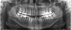
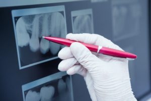
The panoramic x-ray allows the dentist to diagnose and study various problems of the oral cavity, such as bone abnormalities, teeth and jaw fractures, impacted teeth, types of periodontal disease, cysts, tumors, etc. The panoramic radiograph is also a valuable tool for implant treatment planning, as well as in the orthodontics.
Its disadvantage though, is that it lacks high resolution.

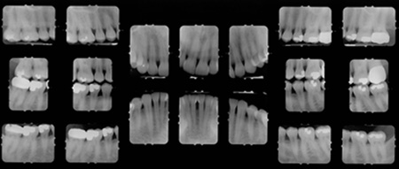
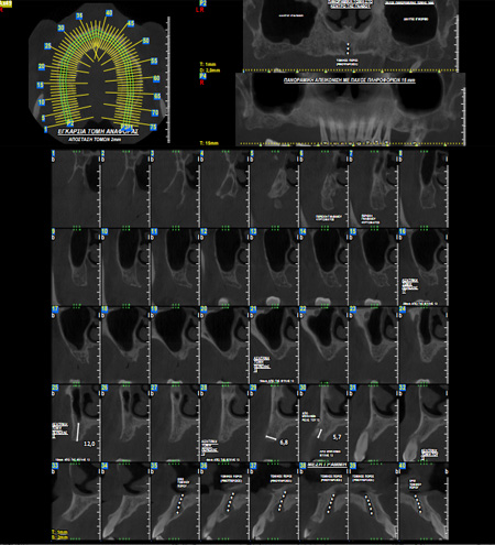
It provides the highest resolution of all the dental radiographs. However, its cost is especially high, as compared to other types of x-rays, and therefore is taken only when necessary.
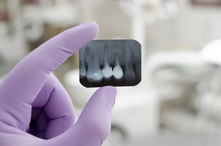
Are dental radiographs safe?
Nowadays, the use of dental radiographs is safe. The radiographic machines, and especially the digital ones, radiate a small dose of radiation. For instance, the radiation received with a panoramic x-ray, is equivalent to the radiation that one receives while watching television for a few hours. The high tech machines also, provide the ability to adjust the magnitude of radiation, depending on the body type and the age of the patient. Thus, radiation is the smallest possible. For extra protection, patients wear, also, special collars and lead aprons that protect other parts of the body from the exposure to radiation.

Use of lead apron
Is it allowed to take dental radiographs during childhood?
Taking radiographs during childhood is allowed, but effort is placed to minimize them to the absolutely necessary. For bigger protection of the little patients, the dose of radiation is reduced, according to their body type and age. Moreover, like all patients, they wear special collars and lead aprons that protect other parts of the body from the exposure to radiation. Finally, because of their dimensionally small mouth, in the case of intraoral periapical x-rays, smaller radiographic tiles are used.
Is it allowed to take dental radiographs during pregnancy?
During pregnancy, it is generally recommended to avoid taking radiographs, unless it is absolutely necessary. Those women, like all patients, wear special lead collars and aprons that protect other parts of the body from the exposure to radiation.
What are the differences of a digital and an analogical radiograph?
Nowadays, the number of dental offices that take digital x-rays is increasing. This shift is due to the advantages of the digital radiographs such as:
- smaller dose of radiation is needed
- faster exposure
- no need for development fluids
- they are easily saved and transferred because of their digital form
How often should I take a panoramic radiograph?
The necessity of a new panoramic x-ray is determined by the dentist, by co-evaluating the clinical condition of the patient, the patient’s age and the confidence of the dentist that the new radiograph will provide new information.
Generally, even though the modern radiographs are safe, their shooting has to be justified and to comply with all protection measures for the patients and the dental personnel.